| Keynote lecture |
| "Nuclear Imaging in the Realm of Medical Imaging" |
In medical imaging, information concerning the anatomy or biological processes of a patient is detected and presented on film or screen for interpretation by a reader. The information flow from patient to reader optimally implies:
* the emission, transmission or reflection of information carriers, typically photons or sound waves, which have to be correctly modulated by patient information through interactions in the patient
* their detection by adequate imaging equipment preserving essential spectral, spatial and/or temporal information
* the presentation of the information in the most perceivable way
* the observation by an unbiased and trained expert.
In reality, only an approximation to this optimal situation is achieved, but it is the ultimate goal of R&D in the medical imaging field to approach the optimum as much as possible within societal constraints such as patient risk and comfort, economics etc...
In nuclear imaging, traces of an appropriate radioactive pharmaceutical are administered to a patient. The radiotracers decay by emitting gamma rays or positrons that can be directly or indirectly detected. The distribution of the radioactive tracers, which is assumed to model information about the patient's anatomy and physiology, is then inferred from the detected gamma rays and mapped as a function of time and/or space.
Radionuclides such as Tc-99m decay by emitting single gamma rays. These rays can be detected and the original tracer distribution can be directly visualised as projections by means of a gamma camera or SPECT camera. By measuring projections over an adequate set of angels (p or 2p), tomographic reconstruction techniques can be applied to generate images of the tracer distribution in chosen virtual slices of the body.
When the decay process generates a positron, such as with F-18, the positron travels in the tissue for about 2 mm before colliding and annihilating with an electron. The energy equivalent of both the electron and positron mass is then liberated in the form of two oppositely directed coincident gamma rays of 511 keV. By detecting a large number of coincident photons over enough angles, tomographic reconstruction techniques yield images of the original radionuclide distribution, using either a dedicated positron camera or a double headed gamma camera in coincidence mode.
The images thus obtained only represent approximations of the true tracer distribution. Indeed, the interactions of the photons with the medium in which they originate and travel will alter the information they may carry. Many photons will disappear during the imaging process through absorption. Others will change direction and energy due to scattering. Only a small percentage of the emitted photons will eventually contribute to the image and even less of them will carry information which improves the final image quality in view of the original diagnostic goal. Some image processing techniques may reduce or overcome limitations in the imaging process.
We will first introduce the basic physical concepts underlying the imaging process. We will then shortly situate different imaging modalities in the realm of medical imaging with some emphasis on nuclear imaging.
|
Frank Deconinck
Nuclear Medicine VUB
Laarbeeklaan 101
B-1090 Brussels, Belgium
Tel +32 2 477 46 10
Fax +32 2 477 46 13
frank.deconinck@vub.ac.be |
 |
Frank Deconinck studied physics at the Universiteit Gent and the Vrije Universiteit Brussel (VUB). He obtained his PhD in 1977 on medical applications of semiconductor detectors. After working at the University of California in San Francisco and at Brookhaven National Laboratory he became professor of medical physics at VUB. His research activities are mainly in the field of medical image processing and in access to visual information by blind people. Together with his colleague A.Bossuyt, he obtained the Hewlett-Packard prize for medical informatics (1984) for his work in nuclear cardiology.
Next to his activities at VUB he was chairman or vice-chairman of the Belgian Nuclear Waste Agency NIRAS-ONDRAF, of the Belgian nuclear waste treatment plant Belgoprocess, and of the Governmental Agency for Non-Proliferation of Atomic Weapons CANVEK. He is currently chairman of the Board of Governors of the Belgian Nuclear Research Centre, SCK•CEN, vice-chairman of Belgonucleaire and chairman of the Belgian Nuclear Society, BNS.
Together with his wife he organised the exhibition 'Tactile Graphic Art', selected by UNESCO for the U.N. World decade for cultural development. Exhibitions were held in Belgium, Paris, Köln, Osaka, Tokyo, ...
He is co-author or editor of 5 books, 90 articles, 180 communications and 50 abstracts.
|
| Keynote lecture |
| "The clinical value of X-ray images of the teeth and jaws" |
X-ray images of the teeth and jaws are required for a variety of diagnostic purposes to supplement a clinical examination, and are invaluable for providing pictorial information about the structures that cannot be examined with direct vision. They can be acquired using traditional film systems which produce analogue images, or using a variety of alternative image receptors which result in a digital image. Irrespective of the method of image acquisition they are utilised by the dental surgeon in order to benefit their patients' management.
Numerous disease processes can affect the teeth and jaws, in addition to a range of developmental abnormalities. These can be classified according to a radiological sieve, which also includes artefactual and iatrogenic features. The key components of the classification are: developmental, inflammatory, traumatic, cystic, neoplastic, osteodystrophies and systemic disorders.
This presentation will utilise examples of radiographic images of patients to illustrate components of this classification which are unique to the teeth and jaws.
|
Laetitia Brocklebank
Glasgow Dental Hospital & School
378 Sauchiehall Street
Glasgow G2 3JZ, UK
Tel. 44 (0)141 211 9640
Fax. 44 (0)141 331 2798
email: l.m.brocklebank@dental.gla.ac.uk |
 |
Laetitia Brocklebank originally qualified in dentistry from Guy's Hospital at the University of London. She undertook her radiology training at the University of London, and in Hong Kong. Since 1991 she is appointed as a Senior Lecturer in Oral Radiology, University of Glasgow, and as a Honorary Consultant in Oral Radiology, Greater Glasgow Health Board, Glasgow Dental Hospital and School, 378 Sauchiehall Street, Glasgow G2 3JZ, UK.
Her research interests are in digital radiology, with an emphasis on reconstruction and subtraction applications. She has been actively involved in developing:
* a technique for standardisation of image geometry when taking intra-oral digital (or film) radiographs
* specialised software for carrying out geometric reconstruction of digital images prior to analysing by subtraction
* specialised software for density adjustment of digital images prior to subtraction
The thrust for this research is to be able to use digital subtraction radiology to assess bone, or tooth change, in a quantitative as well as qualitative manner. Application of this flexible and user-friendly technique would be advantageous in longitudinal evaluation of:
* dental trauma
* periradicular surgery
* periodontal disease and treatment
* implants
She has spoken at many national and international meetings about aspects of interpretation of images of the teeth and jaws and in 1997 published the book: "Dental Radiology: understanding the X-ray image" (Oxford University Press).
|
| Keynote lecture |
| "Intra-oral and extra-oral digital imaging: an overview of factors relevant to detector design" |
Since the introduction of the first intraoral detector system 15 years ago, major improvements have been achieved. Initially fiber optic coupling was used between the entrance phosphor and the small CCD sensor, resulting in distortion of the image. Later sensors became available for direct exposure. Also the dimensions of the active area were increased to comply with the size of analogue dental films. The thickness of the sensor was decreased as well.
Intraoral films are flexible. This property is a drawback in the proper display of the geometrical properties of the object. CCD-type sensors however, are stiff and therefore much more difficult to position in the mouth of the patient.
The resolution of modern intraoral sensors is comparable or even higher than that of film. However, with increasing resolution introduction of noise is becoming a problem in some diagnostic tasks such as the detection of low contrast lesions, e.g. incipient caries.
In extra oral imaging also use is made of both imaging phosphor plates and CCD-based systems. The procedures in Phosphor plate based technology are quite comparable to those with conventional film. As the width of sensors is limited, use is made of a slit imaging technique in panoramic imaging and still images.
Tomography, a radiographic method to display layers within an object is not possible with the present CCD sensors. For these images dimensions of CCD sensors are required which are equal in size to those of the x-ray beam (5x7 cm).
|
Gerard C.H. Sanderink, DDS, PhD
Dept. Oral and Maxillofacial Radiology
Academic Centre for Dentistry Amsterdam
Louwesweg 1
1066 EA Amsterdam
The Netherlands
phone +31 20 5188 260
fax +31 20 5188 480
e-mail G.Sanderink@acta.nl |
 |
Gerard C.H. Sanderink obtained his DDS degree in 1971 at the University of Utrecht and joined the scientific staff of the Department of Oral Radiology of the Faculty of Dentistry. Since 1986 he is a member (Associate Prof.) of the scientific staff of the Department of Oral Radiology of the Academic Centre for Dentistry in Amsterdam.
Panoramic techniques have been one of the research subjects which resulted in a PhD degree in 1987 on a dissertation entitled: 'Imaging characteristic in panoramic radiography'.
His current research interests and speciality include digital intraoral and panoramic systems, digital image processing and image quality assessment in clinical dental radiology.
Another main interest is the development and introduction of Computer Aided Instruction in the educational programme of the Dental School. At present he is chairman of this group and responsible for implementation of it in the curriculum.
Since 1980 he has been active in the International Association on Dento-Maxillo-Facial Radiology (IADMFR), as Vice President (1980-1985) and 1997 he was elected as Secretary General.
|
| Keynote lecture |
| "Detectors for imaging biological macro-molecules at atomic resolution" |
Knowledge of the atomic details of biological macro-molecules is required for understanding life's fundamental processes. This information is essential not just to satisfy our curiosity, but also for the design and development of new drugs to combat disease.
Atomic details in biological macro-molecules can be visualized by X-rays and electrons. Crystal diffraction is required for visualization by X-rays, whilst biological macro-molecules can be visualized through imaging and diffraction by electrons. The ideal detectors will be described for these three different modes of imaging and an overview of current detectors will be given. On the basis of these data, a summary of the next challenges in detector development will be formulated.
|
prof. Dr J.P. Abrahams
Biophysical Structural Chemistry
Leiden Institute of Chemistry
P.O. Box 9502
2300 RA Leiden, The Netherlands
tel +31 71 5274213
fax +31 71 5274357 |
 |
| Jan Abrahams received his Ph. D. Degree, cum laude, for his work at Leiden University on the biochemical and biophysical characterization of factors essential for protein synthesis. Subsequently he joined the MRC Laboratory of Molecular Biology in Cambridge, UK. Here he elucidated by X-ray crystallography the structure and mechanism of various proteins and protein complexes, including the structure of F1 ATPase. He showed this protein complex to be a nanoscopic dynamo with stationary and rotating elements. This discovery led to the award of the Nobel Prize to Dr J. Walker, the leader of the research team. In Cambridge he also developed software and hardware for protein crystallography. After seven years in Cambridge he moved to the Leiden Institute of Chemistry to start a group in biophysical structural chemistry. The research group he heads develops methods for nano-crystallogenesis, for structure determination by cryo-electron microscopy and for structure solution and refinement by X-ray crystallography. In addition the group researches fundamental aspects of apoptosis, ion pumping, bacteriophage infection and cell-cell interactions.
|
| Keynote lecture |
| "Flat X-ray Detectors for Medical Imaging" |
Medical X-ray Imaging is currently seeing the introduction of Flat X-ray Detectors. Static Flat Detectors replace screen-film and storage phosphor systems, while dynamic Flat Detectors can be used instead of Image Intensifier Tubes with TV cameras.
Especially for Cardio applications dynamic Flat Detectors systems are entering the market.
Flat X-ray Detectors are based on amorphous silicon large area electronics, the same technology also widely employed in today's flat display devices (TFT displays). Philips has been strongly involved in the research and development of Flat X-ray Detectors for more than a decade. This presentation gives an insight into the principles Flat X-ray Detectors are based on. Also the components used to build such large area X-ray detectors will be discussed. Finally, performance data of Flat X-ray Detectors are presented.
|
Dr. Michael Overdick
Philips Research Laboratories
Weisshausstr. 2
52066 Aachen
Germany
Tel +49 241 6003-567
Fax +49 241 6003-442
michael.overdick@philips.com |
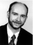 |
| Michael Overdick (born 1969 in Porz am Rhein, Germany) studied Physics at Bonn, Germany and Cambridge, England. He received an MPhil from the University of Cambridge in 1993 for coincidence experiments on secondary electron emission from solids. For his PhD at Bonn University he worked on a digital autoradiography system based on silicon strip detectors. In 1998 Michael Overdick joined Philips Research in Aachen where he is active in the field of flat medical X-ray detectors, especially based on amorphous silicon large area electronics. His main research interests are X-ray conversion, associated temporal effects and general X-ray detector design.
|


